

Tricuspid Valve
The Tricuspid valve is the atrioventricular valve present in the right side of the heart. There are some common features that are present in the atrioventricular valves (i.e. the Tricuspid Valve and the Mitral Valve).
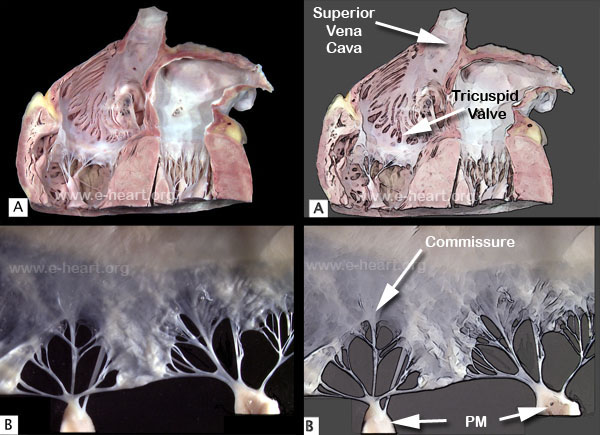
A. Coronal section of the base of the heart showing the atrioventricular valves. B. Posterior tricuspid valve leaflet showing two minor commissures that divide it into scallops. The commissures have corresponding papillary muscles that anchor the fan-shaped chordae. These fine chordae insert to the free edge of the leaflet producing a serrated border. In comparison the Mitral Valve is thicker than the tricuspid valve. However the basic layers of both types of atrioventricular valves are rather similar. The tricuspid valve has a complete annulus or ring which is part of the fibrous skeleton of the heart. The support of the valvular apparatus of the tricuspid valve is not constant, as it shows variable patterns of papillary muscle support
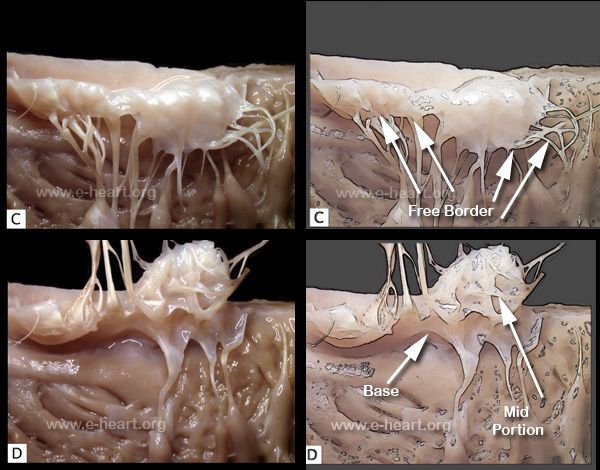
C. The atrial surface of atrioventricular valve leaflets are smooth with variable degrees of ballooning that ensure tight apposition during systole. D. In contrast, the ventricular surface is marked by chordal attachment to different zones of the leaflet. Chordae arising from the heads of papillary muscles can insert on the free edge (first order) or on the ventricular mid zone (second order) of the leaflets. Chordae that originate from trabeculae carneae and insert on the basal zone of the leaflets are called chordae of the third order.
If you want to compare the tricuspid valve to the mitral valve click here.
Back to cardiac structure
.