

Fibrous Skeleton of the Heart
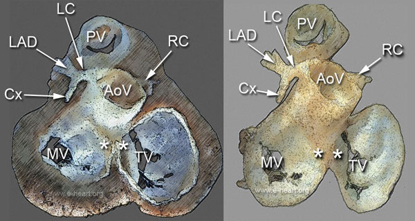 The fibrous skeleton provides attachment for the valve leaflets as well as for the atrial and ventricular myocardium. It consists of the distinct valve annuli and intervening fibrous trigones. The aortic valve (AoV) occupies a central position at the base of the heart and anchors the other three valves. The image on the left is a cephalad view of the base of the heart after removing the atria, epicardial fat and coronary arteries to show the relationships between the atrioventricular and semilunar valves. The mitral valve ring is almost circular and smaller than the tricuspid ring. The mitral valve (MV) shows the anteroseptal or anteromedial and the posterolateral leaflets. The tricuspid valve (TV) has one septal, one posterior and one anterior leaflet. The medial aspect of the tricuspid valve ring merges with the central fibrous body of the fibrous skeleton and the lateral and posterior aspects of the ring provide an anchoring structure to the right atrium. The rigth ventricular free and posterior walls are also anchored to the tricuspid annulus. The aortic root is at the center of the fibrous annulus and is the only structure that connects with all the other three valves. The pulmonic valve (PV) is the most anterior of the valves. There are two trigones, one on the left side and one on the right side, dorsal to the aortic annulus (asterisks). The left fibrous trigone is formed between the anterior mitral leaflet and the aortic valve in the area of the left-posterior aortic commissure.
The panel on the right shows a cephalad view of the fibrous annulus after removal of all the cardiac muscle that was present in the left pane. The tricuspid annulus has a thin lateral border, which according to some authors is incomplete. The left trigone is better appreciated after removal of the myocardium. The coronary arteries (LC, LAD Cx and RC) are clearly shown as they arise from the coronary sinuses in the root of the aorta
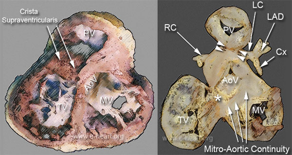 Caudal view of the fibrous skeleton of the heart. The image on the left shows that the inflow and outflow tracts of the right ventricle are separated by the crista supraventricularis (CSV). The anterior leaflet of the mitral valve is seen in continuity with the aortic valve.
The panel on the right shows a caudal view of the fibrous skeleton after removal of all the myocardium. The only muscular structures left in the specimen are the tips of the papillary muscles attached to the valve leaflets. The right fibrous trigone or central fibrous body, which is formed between the anteroseptal tricuspid commissure, anterior mitral leaflet and right-posterior aortic commissure, is shown (asterisk). The pulmonic valve ring is attached to the aortic valve ring by a small conus ligament (arrows). Note that most of the support of the pulmonic valve is provided by the crista supraventricularis, the septal position of the right ventricular outflow tract. Also note that the pulmonic valve has an orientation opposite to that of the aortic valve, due to a criss-cross arrangement of the two outflow tracts.
Back to Cardiac Structure