

Histology of the Pericardium - I
 A. Normal, full thickness parietal pericardium is shown. It consists of a layer of mesothelial cells (dark nuclei on the top surface) and compact layers of dense wavy fibrous tissue (collagen) (yellow) with interspersed scant short elastic fibers (black fine lines between the yellow fibrous tissue bundles).
B. Fibroblasts with elongated nuclei and scant thin blood vessels are normally present in the parietal pericardium. CD34 immunostaining highlights the endothelial cells of capillaries.
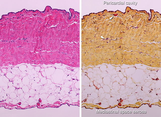 These microscopic images of the lateral wall of the parietal pericardium shows a serosal layer of pericardial mesothelial cells lining the pericardial cavity and the fibrosa layer. The fibrosa layer is the thick eosinophilic layer (left image) or yellow layer (right image) made up of dense wavy collagen fibers. Faint black lines represent a minimal amount of elastic lamellae in the fibrosa. The parietal pericardium contains a few small blood vessels (arrows). Between the fibrosa of the parietal pericardium and the mediastinal parietal pleura is a layer of epipericardial fat. Note the serosal layer of the pleura is also made up of a layer of mesothelial cells. (X50, H&E and Movat pentachrome).
The mid magnification of the histology of the visceral pericardium (epicardium) and the praietal pericardium (epicardium) is shown here.
Detailed view of the Serosal (mesothelial cell) component is shown here.
Back to Pericardial disease
Back to Home Page