

Histology of the Pericardium - III
The serosal component of the pericardium consists of a single layer of mesothelial. The mesothelial cell layer forms the serosal parietal and a visceral layers that line or invest the fibrous (parietal) and visceral pericardium.
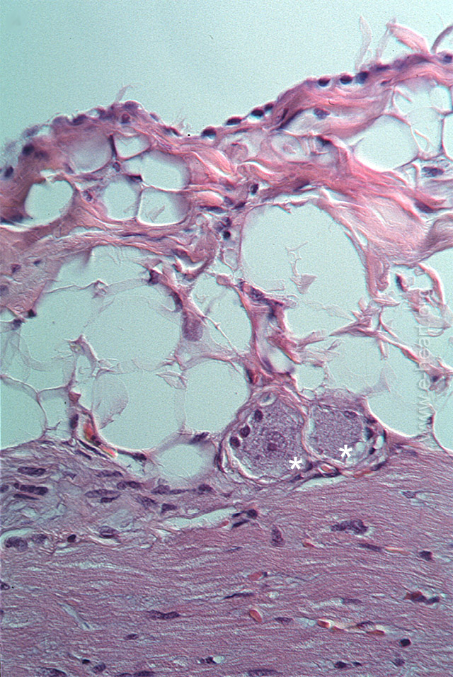
In this image there is adipose tissue on the epicardium overlying the subjacent myocardium. The asterisk (**) illustrate two ganglion cells on the epicardium. In addition adipose tissue is also frequently present on the external (mediastinal) surface of the parietal pericardium particularly on the left anterior cardiophrenic angle.
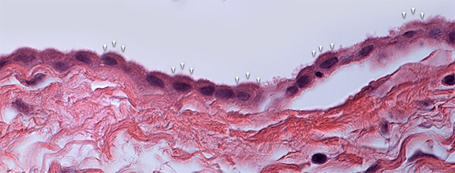
A sheet of mesothelial cells is shown at high magnification lining the fibrosa layer of the parietal pericardium. They are identical to the mesothelial cells seen in the visceral serosa shown above. These cells can vary from flat to cuboidal in shape. Microvilli (arrowheads) are present on the surface facing the pericardial cavity to increase the surface area of these cells. The microvilli impart a fuzzy look to the luminal border of the mesothelial cells.
Back to Pericardial disease