

Normal Pericardial Anatomy - Phrenic Nerves - I
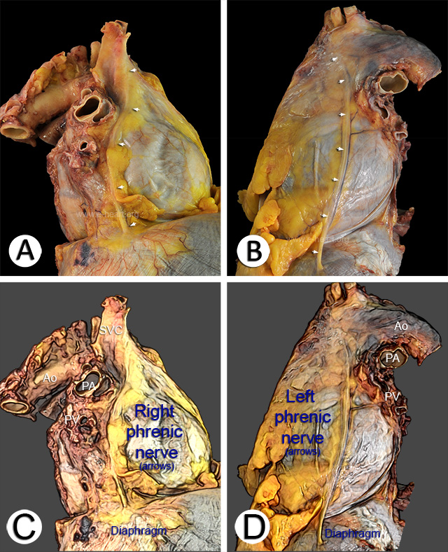 A and B. Right lateral view of the pericardium shows the right phrenic nerve (arrows). It courses parallel and lateral to the superior vena cava and continues downward, anterior to the right pulmonary hilum, towards the diaphragm. C and D. Left lateral view shows the left phrenic nerve (arrows) descending over the left atrial appendage and anterolateral left ventricle before reaching the diaphragm.
Note the relationship of the pathway of the phrenic nerve intersecting with the path of some of the coronary arteries and veins visible through the translucent myocardium. This path can allow pacing devices deployed in the coronary veins to stimulate the phrenic nerve as well, which is an undesirable effect.
Four additional examples of slight variations of the course of the phrenic nerves over the parietal pericardium are shown in the next 4 pages (2, 3, 4, 5).
The close relationship between the parietal (fibrous) pericardium and visceral pericardium as well as the pericardial space are shown here.
Back to Pericardial disease
Back to Home Page