

Normal Pericardial Anatomy - Phrenic Nerves - IV
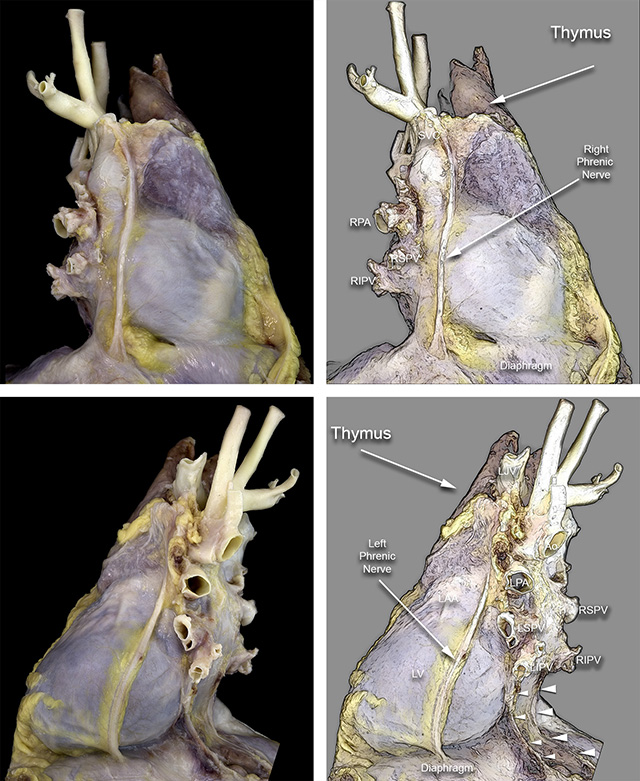
Lateral views of the pericardium covering the heart. The top image is a right lateral view showing the brachiocephalic artery on the uppermost posterior position. Anterior to it the thymus is clearly visible, partially covered by a thin fascia of loose connective tissue. The right phrenic nerve is seen just anterior to the superior vena cava. It follows an almost vertical path on the right border of the heart until it reaches the diaphragm. The lower image shows the left lateral view of the heart and pericardium. The left phrenic nerve is seen emerging just behind the adipose tissue in front of the left pulmonary artery. It follows a roughly vertical direction and moves slightly anteriorly as it courses over the contour of the obtuse margin of the left lateral ventricular wall, and it moves slightly posterior as it reaches the diaphragm.
Ao = Aorta. LJV = Left jugular vein. LPA = Left pulmonary artery. LSPV = Left superior pulmonary vein. LIPV = Left inferior pulmonary vein. RPA = Right pulmonary artery. RIPV = Right inferior pulmonary vein.
RSPV = Right superior pulmonary vein.
The next example shows how the phrenic nerve overlies the coronary veins that drain the lateral wall of the left ventricle. This is of functiontional importance since pacing leads deployed in the coronary sinus and veins of the left ventrcular wall may actually stimulate the left phrenic nerve.
Back to Pericardial disease
Back to Home Page