

Cardiac Structure - Interatrial Atrium
 The upper images show a coronal section of the interatrial septum. The muscular and membranous components components are clearly illustrated. The embryologic septum primum and septum secundum can be seen as discrete muscular walls with intervening connective tissue. The membranous component is the result of fusion and fibrosis of the muscular layers after birth in most human hearts. When these muscular components fail to fuse, the result is a patent foramen ovale. In these instances the right side portion of the fossa ovalis is also called the valve of the fossa. And as long as the blood pressure in the right side of the heart does not increase above the systemic pressure, the foramen ovale remains closed.
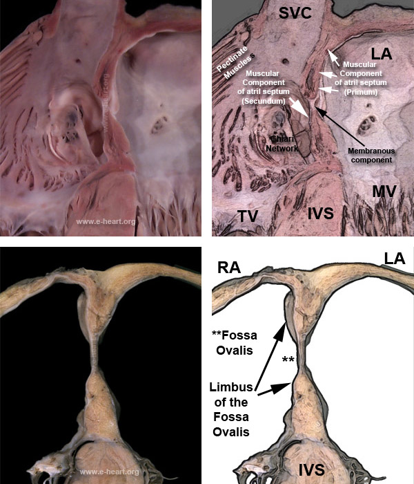
In the adult heart the center of the fossa ovalis is truly a fibrous membrane. The limbus of the fossa ovalis is muscular. However there is usually mesenchymal tissue trapped between the muscular components of the septum. These mesenchymal components can proliferate and form adipose tissue aggregates that thicken the septum. These adipose tissue infiltrates can grow in a significant way and form masses which can be erroneously diagnosed as tumors. This type of thickening secondary to adipose tissue is known as lipomatous hypertrophy of the interatrial septum and does not really represent tumoral growth. It is common in obese patients.