

External Anatomy of the Heart (I)
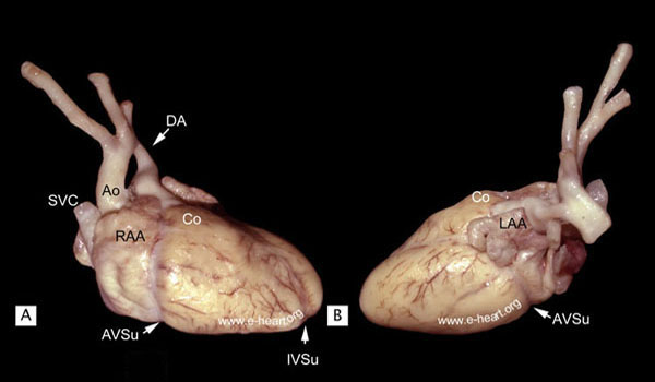
A. Lateral view (right to left). The right atrium is slightly cephalad to the right ventricle. Within the chest, the right atrium is a right lateral chamber while the left atrium is a midline posterior chamber. The right ventricle is anterior and caudal to the right atrium and ventral and to the right of the left ventricle. The right atrial appendage extends anteriorly and partially surrounds the root of the aorta, with which is in intimate contact. The atrioventricular sulcus (AVSu) is clearly demarcated and separates the atrium from the ventricle. The left circumflex and the right coronary arteries course in this sulcus. The superior vena cava (SVC) is seen entering the heart in the far left side of the specimen. The most cephalad portion shows the ascending aorta and the aortic arch. The anterior wall of the right ventricle is somewhat flat except for the portion leading to the outflow tract just below the pulmonary trunk, which is somewhat round and bulging, forming the conus (Co). Note the ductus arteriosus (DA), which communicates the pulmonary trunk with the aorta. B. Left lateral view of the heart shows that the left ventricle is anterior and caudal to the left atrium and posterior and to the left of the right ventricle. The left atrial appendage (LAA) is prominent and shows a very distinctive scalloped border. The atrioventricular sulcus is also distinct. Note the virtual absence of grossly discernible epicardial fat, which makes the branches of the coronary arteries appear relatively more prominent than in the adult heart. In this view, the superior border of the heart is formed by the anterior wall of the right ventricle and the pulmonary conus. Anterior and posterior views are shown here