

External Anatomy of the Heart (II)
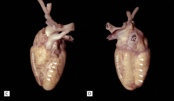
A. Anterior and posterior views. More commonly, the excised heart is depicted with the apex oriented straight downwards, as in these views, to show the four chambers in one plane. The ventricles are demarcated externally by the anterior and posterior interventricular sulci (arrows), over which the anterior and posterior descending coronary arteries follow their course. The two sulci meet at the apex, sometimes forming a notch, as in this specimen. The crux of the heart, located on the posterior aspect, is the point where the posterior interventricular sulcus meets the atrioventricular sulcus. The latter marks the level of the atrioventricular valves. The interventricular sulci correspond to the location of the interventricular septum. These landmarks are very helpful in the external assessment of the sizes of the different chambers in congenital heart disease. In the adult heart these features are less obvious because of the accumulation of adipose tissue on the surface of the heart .