

Atrial appendages in the fetus
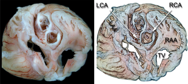 This cephalad view of a 18.5 week old fetal heart with the atria "unroofed" shows the mitral and tricuspid valve (TV). The great vessels are nestled in the middle of the atrial anteriorly. With the aorta being dorsal or posterior to the pulmonary artery. Flanking the great vessels are the atrial appendages. The left atrial appendage (LAA) lumen is narrower than the lumen of the right atrial appendage (RAA). Both have trabeculations (pectinate muscles). The coronary arterial ostia are distinctly visible as they arise from their respective left and right coronary sinuses. In turn, the corresponding left coronary (LCA) and right coronary (RCA) arteries arise from these ostia. The fetal appendages are distinctly defined in the fetus at this age in ventral (anterior) and dorsal (posterior) as well as , anterolateral views.