

Normal Pericardial Anatomy - Parietal & Visceral Pericardium - I
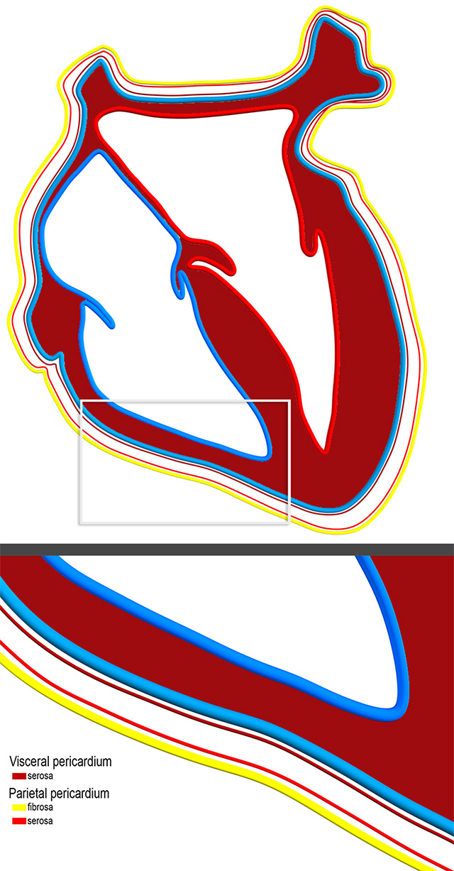 This diagram shows the parietal vs. visceral pericardium. The pericardium has, as many other serosal surfaces, a parietal and a visceral component. The parietal pericardium is composed of two layers: a serosal lining (thin red line) and a fibrous sac (thicker yellow line). The visceral pericardium or epicardium is composed of a single layer of serosal investment covering the entire heart (thin red line overlying the myocardium in blue). Note that the serosal lining of the parietal and visceral pericardium is a continuous layer of mesothelial cells. The serosal layer of the parietal and visceral pericardium face each other. The potential space lined by the serosal layers is the pericardial cavity.
A close up of the parietal (fibrous) pericardium and visceral pericardium (epicardium) of the right and left ventricles (white boxes) is shown here.
Back to Pericardial disease
Back to Home Page
Back to Home Page