

Myxoma - light microscopy
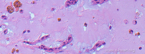 On light microscopic examination the tumor shows an abundant myxoid matrix with spindle cells arranged in short cords and infiltrated by chronic inflammatory cells. Some of the macrophages contain hemosiderin pigment.
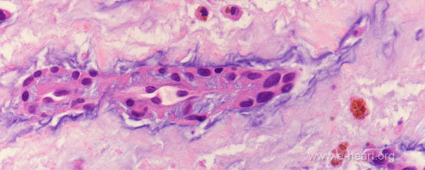 Lepidic cells also called myxoma cells often form ring-like structures around capillaries or form small canaliculi or cords in the myxoid matrix.
 This photomicrograph shows a comparison of the same area of the tumor stained with H&E (left) and stained with a Movat pentachrome stain. The proteoglycan-rich (myxoid) matrix stains green-blue around the cords myxoma ("lepidic") cells.