

The Heart in the Mediastinum
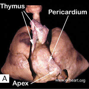 This image shows the heart in the mediastinum with an intact pericardium and the thymus overlying its cephalad portion. Grossly, the pericardium is a roughly conical sac that envelops the heart and the root of the great vessels in the mediastinum. It is attached inferiorly to the tendinous portion of the diaphragm and anteriorly to the sternum through the pericardiosternal ligaments. Posteriorly and superiorly, it is continuous with the adventitia of the arteries and veins of the heart. The pericardiovertebral ligaments provide attachment to the vertebral column. In newborns and young children, the thymus is located in the anterosuperior compartment of the mediastinum and overlies the pericardium, the aortic arch and its branches, and the left brachiocephalic vein..
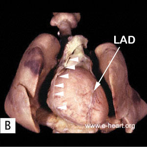 Here the thymus has been removed and a window has been cut in the anterior portion of the pericardium (arrowheads) to show the heart within the pericardial sac. The long axis of the heart normally is directed anteriorly, inferiorly and to the left. Most of the anterior surface of the heart corresponds to the right ventricle; the apex and most of the lateral border, to the left ventricle. The interventricular sulcus (groove) is the place where the left anterior descending (LAD) coronary artery courses through, on its way to the apex of the heart.
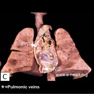 The dorsal portion of the pericardium is revealed after removal of the heart. The openings of The vascular structures entering the pericardium are shown as follows: Ao = Aorta; PA = Pulmonary Artery; SVC = Superior Vena Cava. The asterisks indicate the openings of the pulmonary veins.