

Tricuspid Valve - Cephalad View
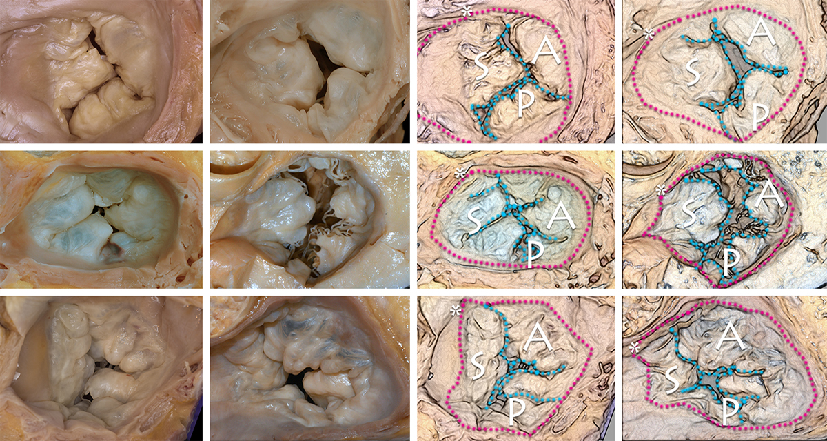
This cephalad view of the six different tricuspid valves illustrates the conspicuous variability in the scallopping patterns of the leaflets. The illustrations on the right correspond to the photographs on the left side of the panel. The red dots roughly delineate the contour of the tricuspid annulus. The white asterisks indicate the begining of the crista supra ventricularis (which would be in anterior-superior anatomical location in the heart in situ. Thus indicating the boundary between the septal (S) and anterior (A) leaflets.
Note the variation in number of scallopings and the depth of the scallopings particularly in the anterior and posterior leaflets. In many instances a discrete posterior leaflet is difficult to discern.
The coronary sinus, if visualized during imaging studies, is a very useful boundary to delimit the extent of the septal leaflet (between the coronary sinus and the crista supraventricularis. Underneath the anterior leaflet in these images is the location of the parietal band of the trabecula septomarginalis.
Back to cardiac structure