

Pulmonic Valve
 The pulmonic cusps are similar in appearance, with three radial, almost symmetric commissural lines of apposition (the linea alba (arrowheads) is a C-shaped thickening of the leaflet near the line of closure of the cusp. The cusps are more or less equal in size and are thinner than the leaflets in the aortic valve.
The collagen bundles that make up the valve leaflet are organized with angular orientation with respect to each other resembling a basket weave. This is very clearly illustrated in the image on the left
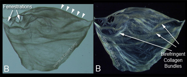 B. Transillumination of a dissected cusp from a young adult. One half of the linea alba of this leaflet is marked by the arrowheads. Fenestrations are commonly seen in the lunulas, but are not hemodynamically significant because the lunulae of adjacent cusps become apposed during valvular closure. The image on the right was photographed while examined using polarized light. The leaflet shows the birefringent nature of the collagen network of the leaflet. This collagen network forms a “basket weave”, with most bundles running parallel to the free edge of the valve.
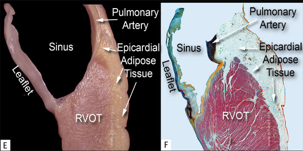 E. View of pulmonic leaflet showing its supporting myocardium and the continuity of this leaflet with the pulmonary artery.
F. Histologic section of the specimen shown in E reveals the supporting muscle. The three layers that form the valve are seen. The mostly collagenous fibrosa (yellow) contrasts with the proteoglycan-rich spongiosa (green) and the ventricularis which has an abundant network of elastic lamellae intermingled with collagen (black and yellow). Also note the transition between the valve cusp and the most basal portion of the pulmonary artery, This zone is rich in collagen (yellow) and devoid of elastic lamellae. (Movat pentachrome satain, 4 X).
Back to cardiac structure