

Cardiac histology (III)
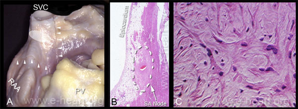 Conduction system. A. The sino-atrial node (SA node) is located at the junction of the superior vena cava with the right atrium along the sulcus terminalis, an external ridge corresponding to the crista terminalis internally. It is roughly elliptical in shape and is located cephalad to the crista terminalis (hatched area). The sinus node artery arises from the right coronary artery in about 55 percent of individuals and from the left circumflex artery in 45 percent. The artery courses through the node and serves as a useful gross landmark for the location of the sinus node. The landmarks of the block of tissue to be cut for obtaining sections of the SA node have been scored with a scalpel (arrowheads). B. This micrograph shows the sinus node. The node is the pale-staining muscle tissue surrounding the central artery (the SA node artery). In contrast, the atrial working myocardium shows a darker eosinophilia. Thus, the SA node is oblong and it is commonly insulated from the working myocardium by fibrous and adipose tissue. (H&E, 4X). In older patients, larger amounts of collagen deposition, loss of pacemaker cells, fatty infiltration and even amyloid deposits can be seen. C. The myocytes of the sinus node are thin and irregular in shape and measure 5 to 6 micrometers in diameter. Cross-striations are also fewer within these cells. They have irregular branching and are arranged haphazardly in an abundant stroma that contains collagenous and elastic fibers. (H&E X 400)
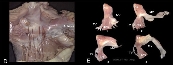
D. The atrioventricular node (AV Node) is located within the triangle of Koch. The anatomic landmarks or this triangle are: 1) the coronary sinus (CS) posteriorly; 2) the line of insertion of the septal leaflet of the tricuspid valve and 3) an oblique line joining these two landmarks. In this specimen parallel serial sections have been made which are perpendicular to the line of insertion of the septal leaflet of the tricuspid valve. The incisions extend from the atrial septum well into the interventricular septum, to sample the AV node. (SVC - superior vena cava, FO - foramen ovale, PV - pulmonic valve ) E. The morphology of the slices from the atrioventricular tissue block is fairly typical. The atrial septum, the tricuspid valve and the mitral valves can be easily recognized for orientation. The insertion of the mitral valve into the fibrous annulus is always more cephalad than that of the tricuspid valve. The most anterior slice should always include the fibrous portion of the interventricular septum close to the posterior cusp of the aortic valve.
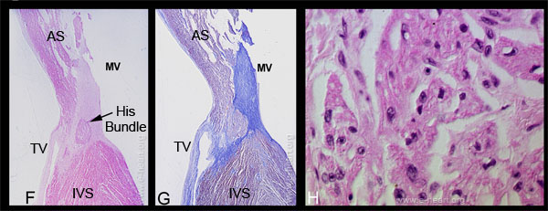 F. Histologic section of slice no. 2 (illustrated in E) shows the proximal-central junction of the AV node as it penetrates the central fibrous body and becomes the His bundle. It courses along the base of the membranous interventricular septum. The site of attachment of the mitral valve to the central fibrous body is labeled MV (H&E 4X). G. The trichrome stain demonstrates the AV node-His bundle tissue as it approaches the fibrous trigone (Masson trichrome 4X). H. Histologic section of the AV node of an adult individual shows that the myocytes are thinner and paler than the working myocytes. Their organization resembles that of the SA node myocytes.
Now that you have learned about the basic cellular components of the cardiac muscle, is time to learn about other very important structures which are part of the heart such as the valves, the conduction system (which is specialized cardiac muscle) and other cellular components.
First, you will learn about the histology of the valvular tissue. This tissue needs constant renewal, since it undergoes continuous motion, with stress to the entire leaflets and trauma to free borders of the valves.