

Cardiac histology (I)
The structure of the heart undergoes constant adaptation to physiologic changes in the organism. These changes are varied and take place from the time of embryonic development to senescence. The difference between physiological and pathological adaptive mechanisms is sometimes subtle. Therefore, it is important to understand that the structure of the heart is modified by a continuum of evolving adaptation of the organ to the needs of the organism. In many instances, pathologic changes affecting the heart have a structural substrate, whether they are due to genetic or acquired processes. Like any other organ, the heart has cells that contribute to the formation of the stroma (the connective tissue scaffold of the organ) and cells that form the parenchyma (the physiological-working-tissue of the organ, in the case of the heart these are the myocytes). Thus the basic cellular unit of the parenchyma of the cardiac muscle is the cardiac myocyte.
This light microscopic image shows normal cardiac myocytes in longitudinal section (accross the screen). The striations on the myocytes correspond to the sarcomeres (see below and sarcomeres).
The myocytes are somewhat tubular in shape. The width of their sarcoplasm (cell cytoplasm) is between 20 and 30 µm. They display a single centrally located nucleus. The connective tissue stroma invests the myocytees and the capillary endothelial cells. The capillaries are present in a 1:1 ratio with the myocytes.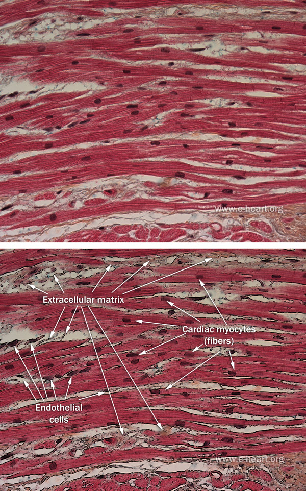
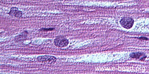
From the histologic standpoint the structure of the cardiac myocyte is best understood if one integrates the knowledge acquired from the study of the basic molecular components of this cell type. The cardiac myocyte is a specialized cell type whose function is to contract. It is striated muscle, which means that when observed through a microscope, the cells of the heart have a banding pattern with periodicity across the length of the cell. At first glance the cardiac myocyte, like any other cell has a cell membrane, which is called the sarcolemma (sarco = muscle; lemma = membrane). The myocyte also has a cytoplasm, which is called the sarcoplasm. It also has a nucleus, which is located in the center of the cell. (Unlike skeletal muscle, in which the nuclei are located towards the periphery of the cell). These two basic components, the sarcolemma and the sarcoplasm of the cell are complex structures that have specialized functions. This image shows typical cardiac myocytes with striations and central nuclei.
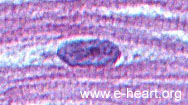 Higher magnification of the micrograph below shows the striated pattern, with a central nucleus (Purple) and a nucleolus (Darker Blue/Red) within the nucleus of the myocyte. The striations are formed by the periodicity of the sarcomeres. The sarcomere is the functional contractile unit of the myocyte. It is composed of several distinct structures. It is delimited by two Z bands. The structure of the myocyte is dynamic, and the cell can grow in size on the basis of physiologic or pathologic stimuli. This growth is called myocyte hypertrophy. In addition, the extracellular matrix supporting the myocytes can also increase, and in pathologic states this is called fibrosis. There are two types of fibrosis in the heart, interstitial fibrosis and replacement fibrosis.
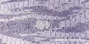 The Z bands or discs are the dark (electron dense) vertical bands seen in the electron micrograph of a myocyte below. The lighter structure in the center is the nucleus. The round darker gray structures around the nucleus are the mitochondria. Thus, this micrograph shows about 8 rows (from top to bottom) of myofibrils composed of sarcomeres (about 19 sarcomeres across the top of the micrograph). The average length of a relaxed sarcomere is 2µm from one Z band to the next Z band.
At higher magnification the detailed structure of the myocyte is shown here.