

Aorta
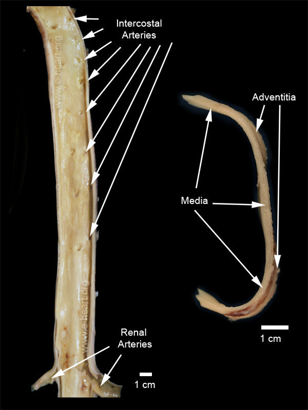 This image illustrates a coronal section of the thoracic and upper abdominal portion of a human aorta from a middle age individual. There are fatty streaks present throughout the surface. There are no complicated atheromatous plaques. The ostia of the intercostal arteries are clearly visible. Since this is the posterior half of the coronal section the celiac trunk and the superior mesenteric arteries are not visible. However the renal arteries are shown.
The image on the right is an enlargement of a cross section of a human aorta. There normal intima is not visible to the naked eye on gross examination. The adventitia is visible as a thin connective tissue layer, which on face shows tiny vasa vasorum.
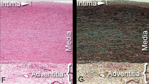 The image on the left side is a hematoxylin and eosin stain of a full thickness histologic section of a human aorta. The intima, media and adventitial layers are clearly demarcated. The intima consists of an endothelial cell layer with two or three subjacent layers of smooth muscle cells without significant elastic laminae.
The media is composed of alternating layers of smooth muscle cells which form circular bundles perpendicular to the axis of blood flow. These layers of smooth muscle cells alternate with elastic laminae. The smooth muscle cells attach to each other and to the elastic lamina. Many connective tissue proteins are present in the normal extracellular matrix of the aortic media.
The adventitia shows a loose connective tissue matrix in which vasa vasorum, nerves and baroreceptors can be found.
The image on the right is a Movat pentachrome stain which clearly demonstrate the elastic laminae (black lines) that alternate with the layers of smooth muscle cells. In the media, the smooth muscle cells stain red, and proteoglycan (mucopolysaccharides) stain green. The adventitial shows yellow connective tissue (collagen).