

Endocardial Lymphocytic Infiltrates -
Quilty Effect - I
Endocardial lymphocytic infiltrates also called Quilty effect lesions. These nodular lesions occur in approximately 10% to 20% of post-transplant endomyocardial biopsies. They may be are confined to the endocardium ( called Quilty type A lesions in the old ISHLT working formulation) or may extend into the underlying myocardium( Quilty type B lesions in the old ISHLT working formulation). However these subtypes have not clinicalc relevacne and thus, they were eliminated in the ISHLT 2004 working formulation. This lesion has been considered distinct from acute rejection, requiring no treatment with intensified immunosuppression. Differentiation of Quilty effect from acute rejection is not usually a problem when the former is confined to the endocardium. However, when it extends into the underlying myocardium, a tangential cut through the biopsy may not show a connection between the myocardial lesion and the endocardial lesion, making differentiation from acute rejection more difficult.20 Cutting additional deeper sections may resolve this dilemma in some cases by demonstrating extension to the endocardium. In the absence of an endocardial extension, the density of the infiltrate, presence of B lymphocytes and plasma cells, background fibrosis and prominent vascularity favor a diagnosis of Quilty effect. Immunohistochemical staining of the infiltrate for B and T cells may be helpful in this regard.
These two images are examples of ELI lesions. The image of the left shows the endocardial location of a small noduele of lymphocytes. The image on the right shows a larger nodule of lymphocytes well contained within the endocardium and a small venule in the center of the lesion.

Lymphocytic infiltrate contained within the endocardium and extending into the myocardium is shown in this biopsy. 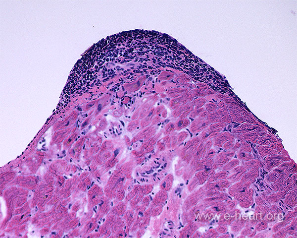
This frozen section shows a Quilty lesion distinctly contained within the endocardium and does not penetrate the subjacent myocardium. Small vessels are commonly present in Quilty lesions (but are more prominent in formalin fixed paraffin embedded tissue. 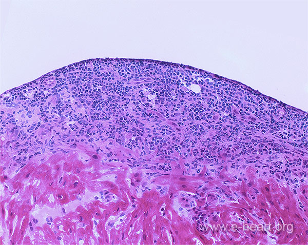
Other examples of ELI lesions are shown here.