

3D Visualization
 Molecules
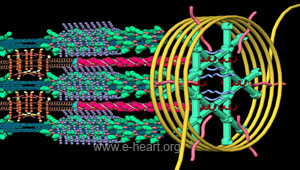
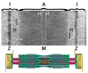
This image is a model that illustrates the proteins that compose the thin and thick filaments, the M band and the Z-disc of the cardiac sarcomere. Higher resolution and details of the organization of the sarcomere are illustrated in the sub-section of molecular anatomy in the Cardiac Structure.
For an animated sracomere beating (contraction of the myosin thick filament pulling the actin thin filament) click here to go to the animated sarcomere page.
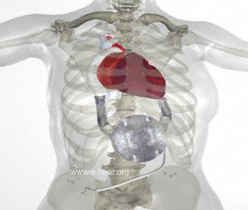
 Assist devices and prosthetic components (valves, stents, counduits, filters, etc.) frequently seen in the practice of cardiovascular pathology can be found in their respective pages.
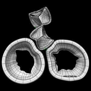
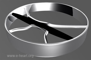
 Structural components of the heart such as the fibrous skeleton or the sarcomeres are easily understood if their 3d structure is taken into account.
In addition other pages dedicated solely to 3D modeling and animation can be browsed here.