

3D - Thin & Thick Filaments & M Band
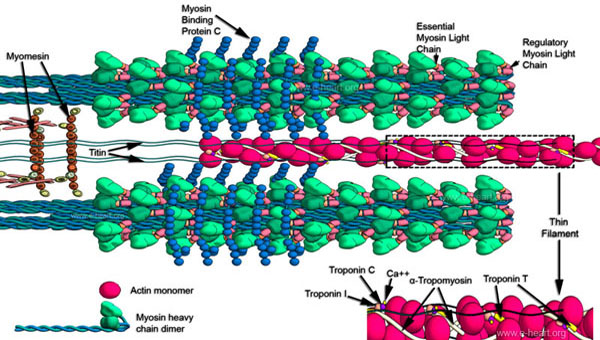
The thick and thin filaments. Myosin (left lower corner) is the monomer that forms the thick filament. Myosin heavy chains form a duplex through their rod domains. They have a "hinge" region which bends and at the end of this hinge region is the myosin head which interacts with actin. The light chains of myosin (regulatory [pink] and essential [orange] chains) are located near the hinge region. The thick filament is made of multiple dimers of myosin (left lower corner) which are organized by overlapping in a helical fashion around the long axis of the filament. The most medial portion of the thick filament consists mainly of the rod portion of multiple dimers which "insert" in the lattice formed by myomesin and other molecules in the M band (dark orange). The myosin binding protein C is represented by the blue spherical monomers. Between the thick filaments, there is a one thin filament (red) illustrated. This is composed of monomers of actin (red spheres) and a helical coil of α-tropomyosin. The troponins (I, C and T) from clusters spaced along the course of the α-tropomyosin. Running parallel to the thick and thin filaments is a giant protein appropriately named titin which spans the entire length of half a sarcomere (about 1 µm). This protein is believed to play a role in sensing stretch. The thin filaments are anchored in the Z band, and a detailed view of the molecules present at this junction is shown here.