

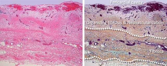
Pericarditis - Organizing - III
Organizing pericarditis with conspicuous neovascularization. Note that the fibrosa layer does not have very conspicuous vessels compared to the area of organizing fibrosis. (H&E stain and Movat pentachrome stain).
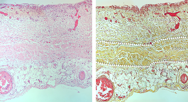
 Maturing organizing pericarditis. Loose edematous granulation tissue becomes organized into denser, thicker fibrous tissue as the amount of extracellular matrix and inflammation diminishes. Relative abundance of fibroblasts (fibroplasia) and newly-formed blood vessels are indicators of the activity of the process.
Denser fibrous tissue forms in organized fibrous pericarditis.
Back to Pericardial disease