

Pericarditis - Organized - II
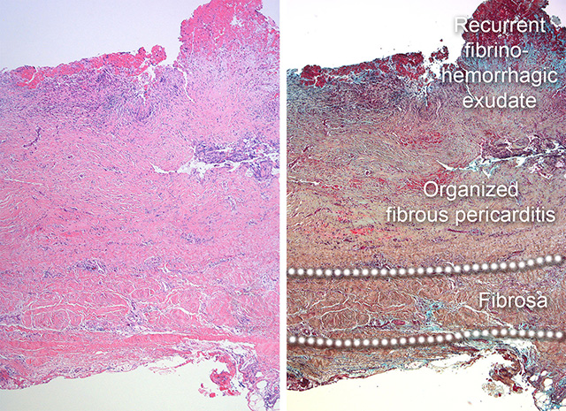
Organized pericarditis with recurrent insult. These micrographs show dense, mature bundles of fibrous tissue overlying the fibrosa layer of the pericardium. The neovascularization is less conspicuous than in the earlier stages of organization. In this example, there is increased cellularity in the upper strata of the pericardium towards the pericardial cavity. This innermost area towards the pericardial cavity shows abundant fibroblast proliferation or fibroplasia, shown as dark blue infiltrates in upper left area of the images. In addition, there is also fibrin and hemorrhage. This is a typical example of a recurrent process with further thickening of the pericardium. Note the original fibrosa layer compared to the excess fibrous tissue, healed and further inflamed. (H&E stain and Movat pentachrome stain).
Back to Pericardial disease
Back to Home Page