Normal Pericardial Anatomy - Pericardial Sinuses and Recesses - III

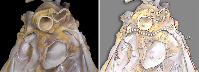
In this anterior view, most of the heart and the anterior portion of the pericardial sac have been removed. Only the dorsal segments of the right atrium, interatrial septum and left atrium remain. The red dots delineate the cut border of the pericardial sac. The asterisks below the atria mark the pericardial space. The white dots delineate the transverse sinus as it spans from right to left. The pericardial space bounded by the cephalad surface of the left superior pulmonary vein and inferior-posterior surface of the left pulmonary is designated the left pulmonic recess (double arrows).
Ao = Aorta. IAS = Interatrial septum. LA = Left atrium. LSPV = Left superior pulmonary vein. PA = Pulmonary artery. RA = Right atrium. SVC = Superior vena cava.
The investment of the ascending aorta by the pericardium forms an aortic recess which can be seen here.
Back to Pericardial disease
Back to Home Page

