

Curriculum - Pericardium
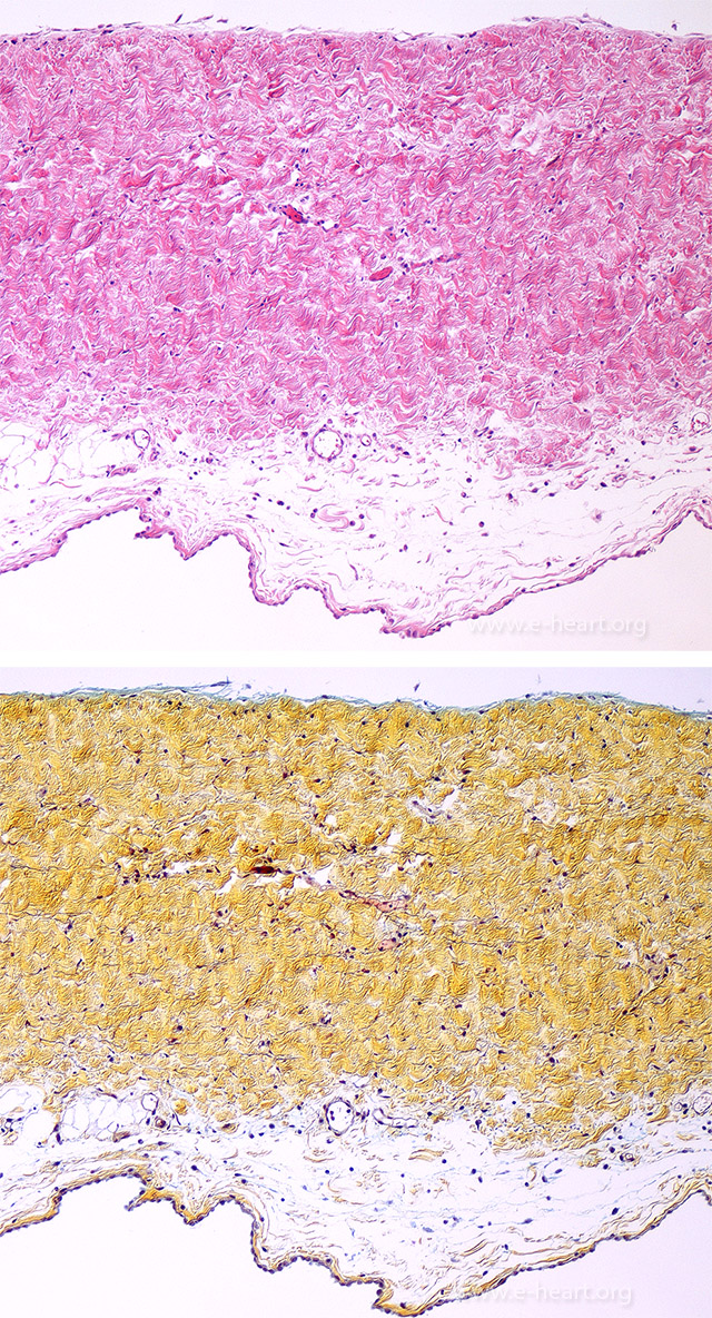 Section of a parietal pericardium showing the fibrous pericardium consisting of compact layers of collagen bundles with a few small vessels and interspersed elastic fibers and serosal pericardium consisting of a layer of mesothelial cells.
 In Fibrinous pericarditis, the strands of fibrin are organized between the parietal (fibrous pericardium) and the visceral pericardium (epicardium). This image shows fibrinous strands over the surface of atrial and ventricles giving the surface a coarse velvety appearance.
 Section of a parietal pericardium with fibrinous pericarditis showing a layer of fibrin with chronic inflammation and inconspicuous mesothelial lining.
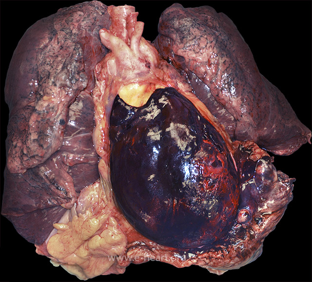 Cardiopulmonary block showing that the anterior portion of the pericardial sac has been removed. This shows the pericardial cavity filled with clotted blood that obscures the heart in a patient who died of cardiac tamponade secondary to a ruptured anterior wall myocardial infarction.
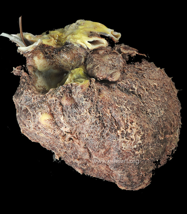
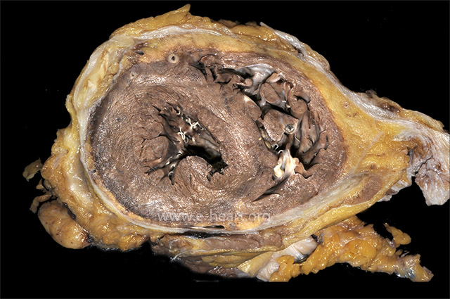 The parietal pericardium is adherent to the heart with obliteration of the pericardial space. There are focal thickenings of the pericardium and calcification in the segment covering the anterior left ventricle.
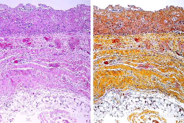
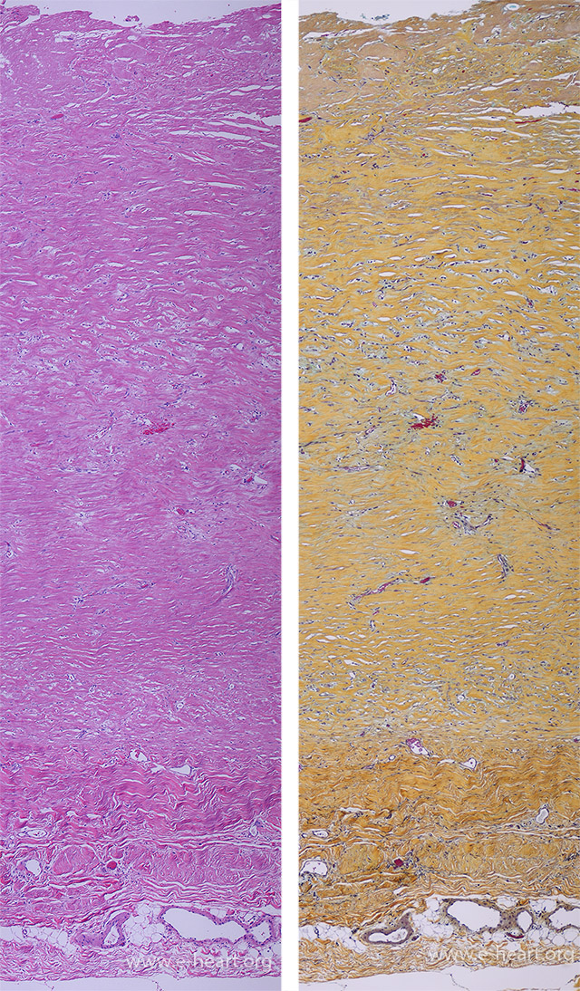 Parietal (fibrous) pericardium (bracket) with marked fibrosis. The dense fibrous tissue is superimposed on the fibrous pericardium. This patient had multiple episode of pericarditis. There are fibroblasts and small capillary sized vessels without inflammation or exudate.
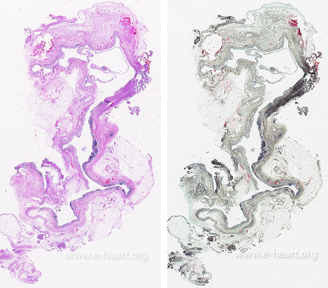 Pericardial cyst are usually unilocular and are lined by mesothelial cells with a fibrous wall and focal aggregates of lymphocytes.
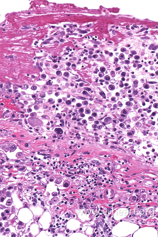 Metastatic adenocarcinoma and fibrinous pericardial exudate in a pericardial biopsy of a patient with recurrent pericardial effusion.