

Curriculum - Tumors of the heart and vessels
 Cardiac myxoma. The images on the left show a myxoma on the surface of a left atrium (the endocardium is thick and white). The tumor is well vascularized, gelatinous and intensely red. A perpendicular section to the atrial surface shows the gelatinous tumor on the endocardial surface of the atrial muscle. The Atrial muscle shows a tan appearance with yellow adipose tissue, indicating that this is interatrial septal muscle. The H&E stain mid panel (top) show a pale myxomatous stroma and in its mid portion is well vascularized and even hemorrhagic. The bottom mid panel shows the matching Movat stain. This stain shows the myocardium in red. The intervening endocardium in yellow and the tumor with its prominent myxoid stroma in green. Higher magnification (right images) show the myxoid stroma with canaliculi of lepidic (myxoma) cells. The Movat stain shows the mucopolysaccharide rich matrix.
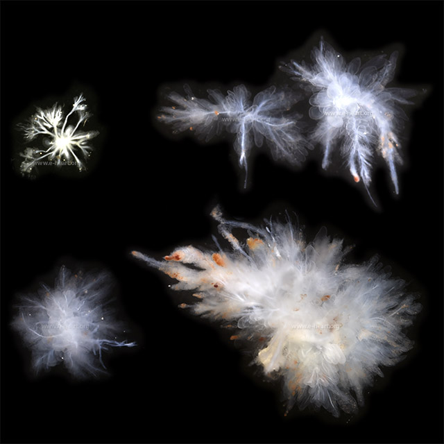 Papillary fibroelastomas usually show a stem from which branches, smaller fronds and actual villi form. The only way to appreciate this architecture is by examining the tumor while immersed in fluid. Commonly there is a swelling of mucopolysaccharide rich matric around individual villi. This gives the tumor an appearance of “bunches of grapes”. In other tumors the villi are just straight. Minute thrombi are trapped between the villi.
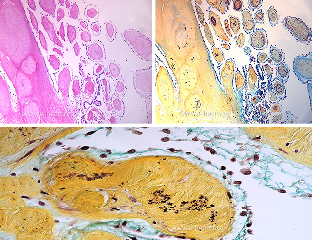 Microscopic examination the papillary fibroelastoma shows the fibrous stem and the braches and villi. The villi show a fibrous stroma shown in the H&E on the left and stained yellow in the Movat stain on the right and lower panel. The fibrous core of the villi shows concentric layers of fragmented elastic lamella (black). The mucopolysaccharide translucent layer shown around the villi in figure 27.62 is shown in the Movat stain as green extracellular matrix.
 Lambl excrescences are morphologically similar to papillary fibroelastomas. They can be single, but commonly they are multiple. They usually do not arborize and do not show a bulging translucent layer surrounding their core. They appear on the free border of the aortic leaflets or on the coaptation border at the level of the linea alba. Less commonly they may be seen the coaptation site near the free border of mitral valve leaflets.
 Rhabdomyoma. The cardiac myocytes have a somewhat polygonal shape and vacuolated appearance with disruption of the myofilaments which imparts a “spider” like branching pattern to the sarcoplasm of the myocytes when seen in cross section.
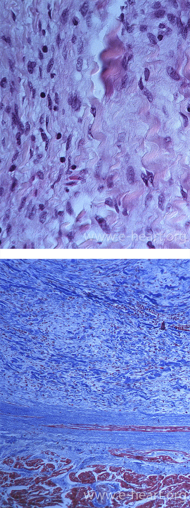 Cardiac fibromas show abundant coarse bundles of birefringent collagen and a conspicuous fibroplasia with slender and plump fibroblasts in the H&E stained upper panel. The tumor is usually well delimited from the myocardium but interdigitations of the tumor fibrous tissue can reach into the myocardial interstitium . The trichrome stain shows the tumor in blue and the myocardium in red.
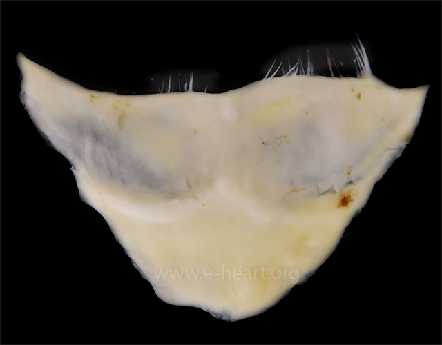
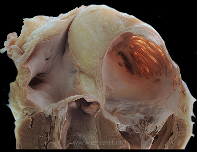 Lipomatous hypertrophy of the atrial septum is shown in this dorsal view of a coronal section through the atria. The two layers of cardiac muscle flanking the adipose tissue are the corresponding layers of septum primum and secundum, which trap mesenchymal cells when they merge and seal. The aortic root (Ao) and valve are seen just caudal to the lipomatous mass.
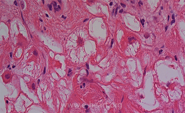
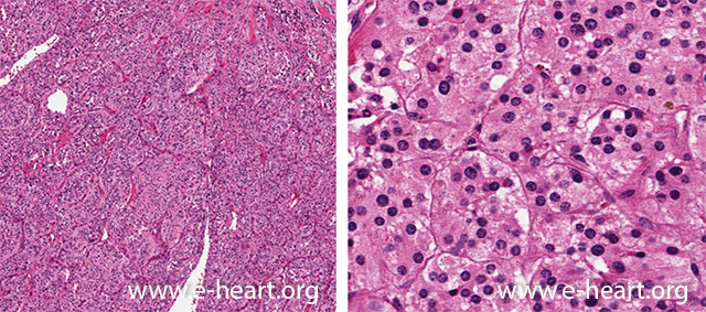 Paragangliomas show the same organoid pattern as they show in any other location. The cells are polygonal with eosinophilic cytoplasm and central nuclei. They form nests which are surrounded by sustentacular cells. Capillaries are conspicuous. The nests of cells are delimited by a thin reticular stroma.
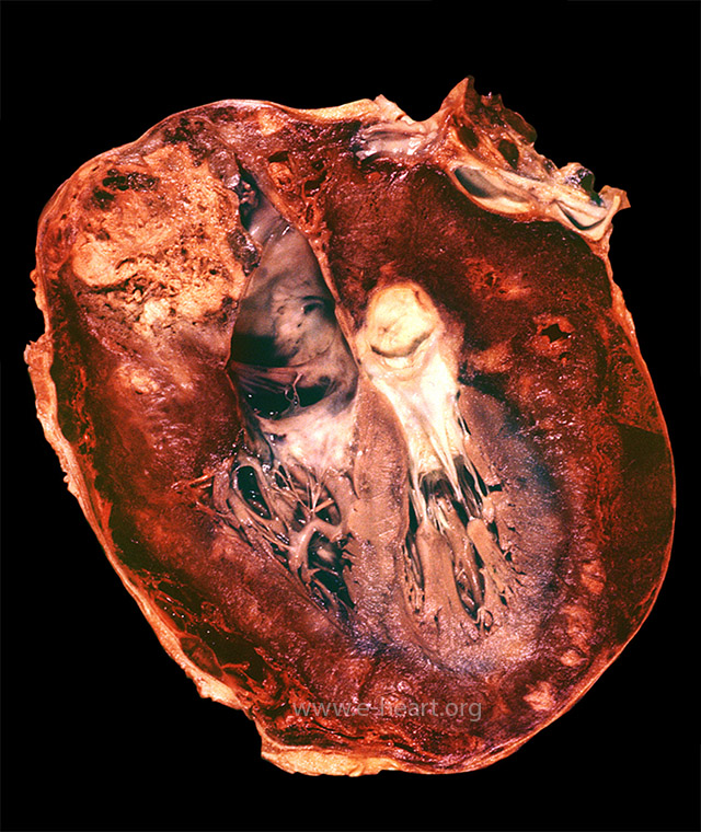 Angiosarcoma of the pericardium surrounds completely the heart and tends to infiltrate the myocardium. It also surrounds the great vessels, venae cavae and pulmonary veins. These features preclude complete surgical removal of the tumor, which often recurs.
 On microscopic examination Angiosarcoma shows channels and papillary structure lined by malignant epithelial cells. Chords forming sinusoids and anastomosing channels are common (upper panel). The malignant cells are positive for CD32 in this image (lower panel).
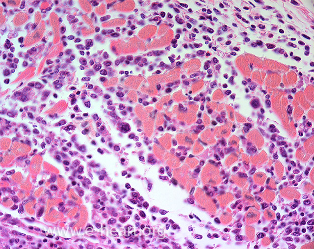 Myocardial infiltration by lymphoma shows conspicuous malignant mononuclear cells in the interstitium between myocytes (endomysium) and between fascicles of myocytes (perimysium).