

Papillary Fibroelastoma
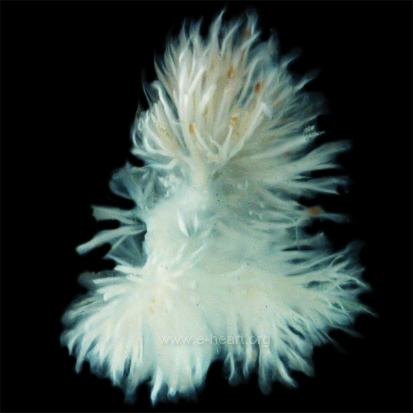 On gross examination the papillary fibroelastoma consists of few to multiple fronds of spindly fronds of white connective tissue. In this specimen there are multiple branches with small spindly papillary structures. Occasionally a stalk is clearly identified.
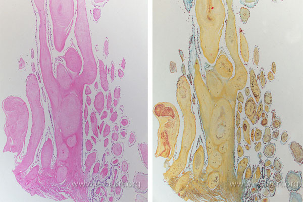 On light microscopy the fronds of connective tissue are eosinophilic (pink) and show minimal cellularity. The most conspicuous cells are endothelial cells on the surface of the "papillary" fronds. The Movat pentacrome (right) shows yellow collagen with concentric, discontinous layers of elastic lamellae in the core. Thus the name fibroelastoma