

Endocardial Lymphocytic Infiltrates -
Quilty Effect - II
This endocardial infiltrate shows a formalin fixed, paraffin embedded tissue biopsy shows early infiltration of the myocardium. In addition, it shows conspicuous blood vessels filled with red blood cells. These are very characteristic and common in Quilty lesions.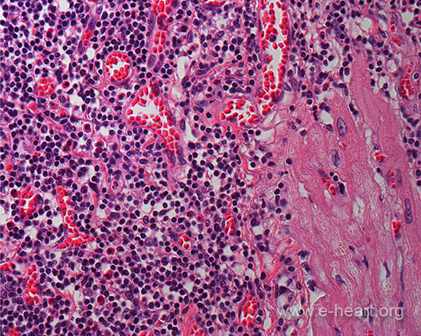


The image on the left shows a thick ELI with numerous small vessels which, in turn, show red blood cells in their lumina. This feature is very useful to identify ELI even in tangential sections.
The image on the right shows a an ELI extending into the subjacent myocardium (Quilty Type B of the old working formulation).
The panel below shows further examples of ELI .
 A. Lymphocytic infiltrate contained within the endocardium and extending into the myocardium is shown in this biopsy. (H&E, X 150). B. This Quilty lesion is distinctly contained within the endocardium and does not penetrate the subjacent myocardium. Such lesion used to be called Quilty type A in the ISHLT-WF-1990. (H&E, X400). C. This infiltrate is clearly within the endocardium and showing early infiltration of the myocardium. In addition, it shows conspicuous blood vessels filled with red blood cells. These are very characteristic and common in Quilty lesions. However in frozen sections, these vessels may not be as conspicuous. (H&E , x 200). D. In this biopsy, the lymphocytic infiltrate clearly extends into the subjacent myocardium with finger-like projections. This used to be called Quilty B in the ISHLT-WF-1990. (H&E, X100)