

Mitral Valve - Rheumatic (I)
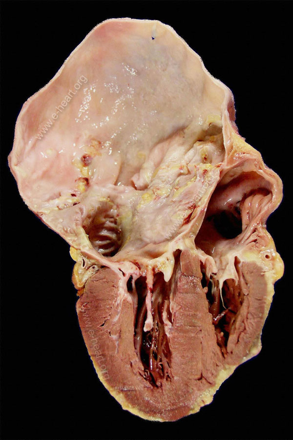 Rheumatic Mitral Valve Stenosis. Dorsal four chamber view of the heart. The left atrium is fibrotic with areas of dystrophic calcification. The mitral valve appears stiff due to severe fibrosis. The fused chordae tendineae are seen as columns of fibrous cord attached to the papillary muscle. The left ventricle is hypertrophied and moderately dilated due to a combination of mitral stenosis and regurgitation. The following image shows a close up of an excised rheumatic valve. Back to valvular disease