

Histologic examination of vegetations remains the gold standard for the diagnosis of infective endocarditis. Histologic criteria for the diagnosis of endocarditis are infected vegetations and valvular inflammatory reaction. Vegetations are composed of fibrin, platelets, neutrophils, necrotic debris and microbial colonies in the acute phase. The valvular inflammatory infiltrates are an admixture of neutrophils, lymphocytes and histiocytes. Rarely, multinucleated giant cells can be found.
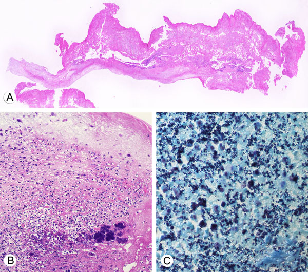
This image shows a microscopic view of native valve endocarditis. The specimen is a mitral valve showing extensive destruction of the leaflet with abundant vegetations (A). The valve is edematous with predominantly polymorphonuclear leukocyte infiltrates and clumps of bacteria (B). The causative organism is Staphylococcus aureus which are seen as Gram-positive cocci (C).
The vegetations in acute endocarditis are practically identical regardless of whether the valve involved is an atrioventricular valve or a semilunar valve . An example of aortic valve endocarditis is shown here.
During indolent infections, there is a predominance of mononuclear cell infiltrates and evidence of neovascularization and fibroblastic proliferation as organization of the vegetation ensues. With chronicity, microcalcifications and fibrosis are formed. The presence of numerous foamy histiocytes should raise suspicion of Whipple's disease. Histiocytes filled with bacteria assume a foamy appearance. Endocarditis caused by intracellular pathogens and fastidious organisms tend to be more fibrotic with calcifications and mononuclear cell infiltrates. Multinucleated giant cells are most commonly observed in viridans streptococcal endocarditis.
Back to endocarditis