

Aortic Valve - Rheumatic (I)
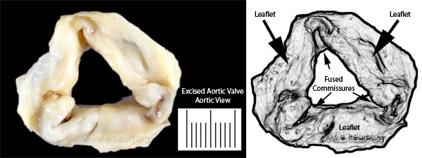 This is a cephalad view of an aortic valve showing three main features 1. There is an overall thickening of the leaflets; 2. There is retraction of the free border of the leaflets and 3. there is fusion of the commissures. The retraction prevents coaptation of the free border (linea alba) of the valve leaflets and creates a central (triangular) orifice that does not close during diastole and thus allow the flow of blood from the aorta back into the left ventricle. In many instances if Aschoff's granulomas are not seen in microscopic examination, it is not possible to differentiate rheumatic disease from other inflammatory conditions that can produce a similar pattern of scarring during healing of the lesion. As a function of time the inflammatory infiltrates disappear and the scarring continues, sometimes to extreme thickening and calcification. Back to valvular disease