

Curriculum - Valvular Heart Disease
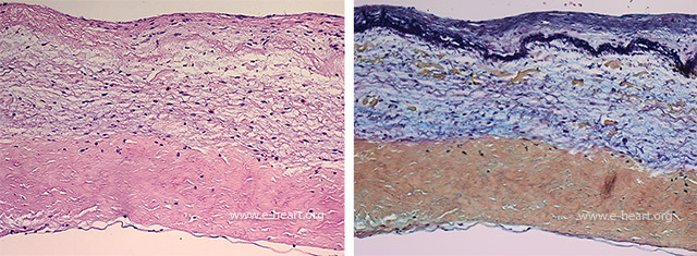 Histology of a normal aortic valve shows three layers as identified from the top to the bottom: ventricularis, spongiosa and fibrosa.
 Histology of a normal aortic valve shows three layers as identified from the top to the bottom: ventricularis, spongiosa and fibrosa.
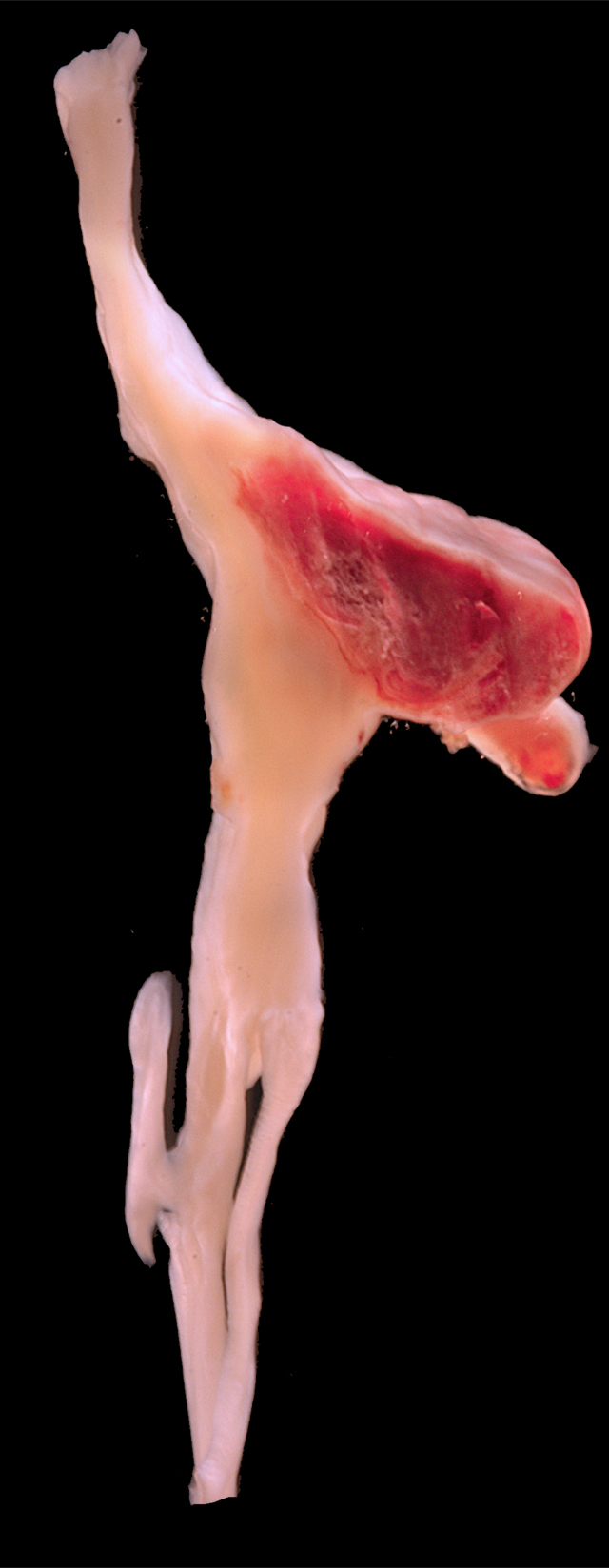
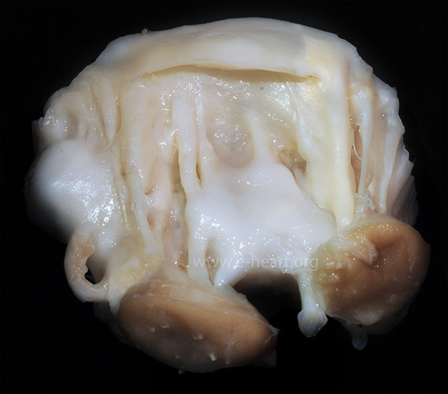 Ventricular view of an excised mitral valve specimen showing shortening of the chordae due to marked fibrosis and fusion. This is a common sequelae of rheumatic valvular disease.
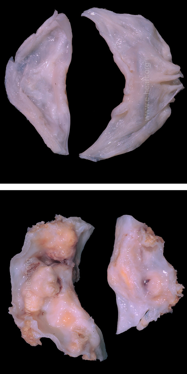 Cephalad view of a bicuspid aortic valve without calcification (Top) shows asymmetry of commissure to commissure distance of the leaflets. A split raphe mimicking a commissure is seen in the cusp on the right. The lower image shows asymmetric leaflets with extensive calcification. The leaflet on the left shows a raphe in the center.
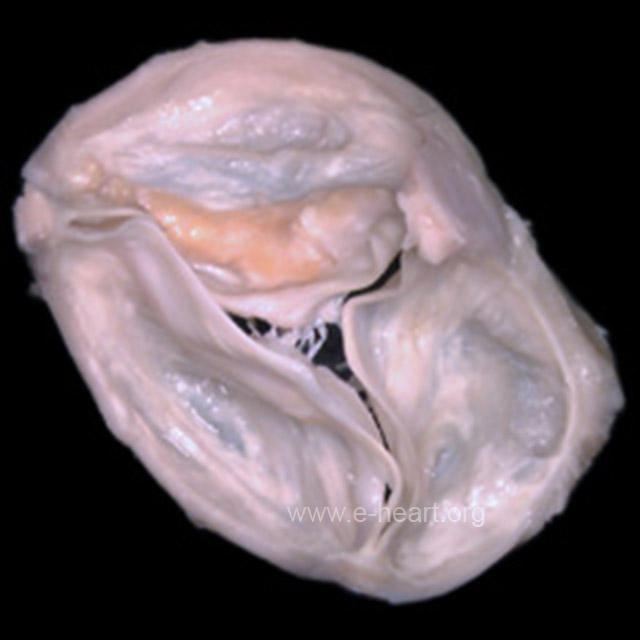 Cephalad view of three-leaflet aortic valve with calcification of a leaflet. The posterior (non-coronary) cusp is (top) shows a yellow area of calcification. The commissures are normal (thin, delicate and non-fused). There are Lambl excrescences protruding from the ventricularis layer of the mid portion of the anatomical right cusp (shown on the left of the image).
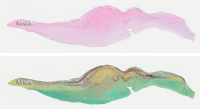 Light micrographs of a tricuspid valve showing a carcinoid plaque encasing the fibrosa (ventricular) layer. The H&E stain shows an indistinct layer of connective tissue on the ventricular surface of the leaflet. The Movat stain (lower image) shows the distinct atrialis layer with abundant elastic fiber (black). The carcinoid plaque is shows proteoglycan rich extracellular matrix (green). Typically carcinoid plaques do not show elastic lamellae. This is a very useful feature to distinguish these plaques.
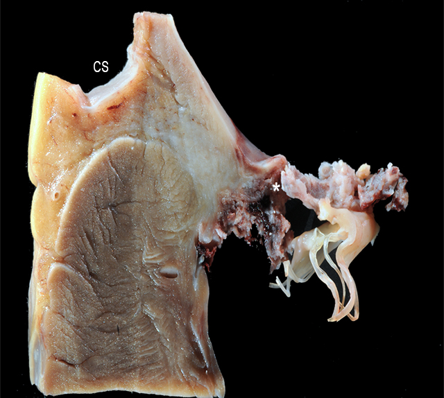 Posterior leaflet of the mitral valve. The coronary sinus (CS) is shown. The mitral leaflet shows fibrinous vegetations on the atrial as well as on the ventricular surfaces. These vegetations communicate through a perforation site on the leaflet (*).
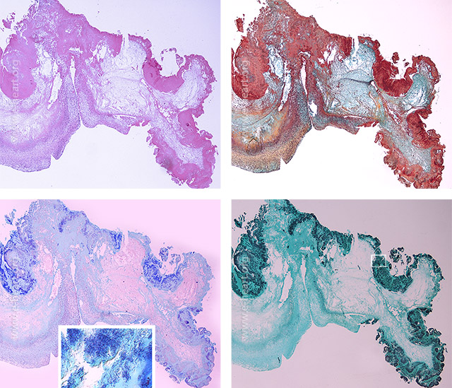 Mitral valve with perforation by fibrinous vegetation. The perforation site is marked by the asterisk. The Movat stain (upper right) shows the loss of continuity of the leaflet at the perforation site. The fibrinous vegetation is shown in red and part of the vegetation shows organization with proteoglycan rich connective tissue (green). The fibrosa of the valve is yellow. The lower panels show the bacterial colonies formed by cocci. Gram stain (lower left and inset) show these cocci. The cocci are also visible as black bacteria in the GMS stain (lower right).
 Posterior leaflet of the mitral valve with a perivalvular abscess extending past the fibrous annulus of the valve into the adipose tissue in the atrioventricular groove. The red dotted line in the lower image delineates the massive vegetation and the abscess with also surrounds the circumflex coronary artery (A) and the great cardiac vein (V) on its path to become the coronary sinus.
 Mitral valve leaflet with a red thrombotic vegetation on the atrial side of the leaflet consistent with non-bacterial thrombotic (marantic) endocarditis.
 The vegetations of non-bacterial thrombotic endocarditis are usually devoid of any significant inflammatory infiltrate in the thrombus. The Movat stain shows the trilaminar architecture of this mitral leaflet and the nil organization of the thrombotic vegetation (red).
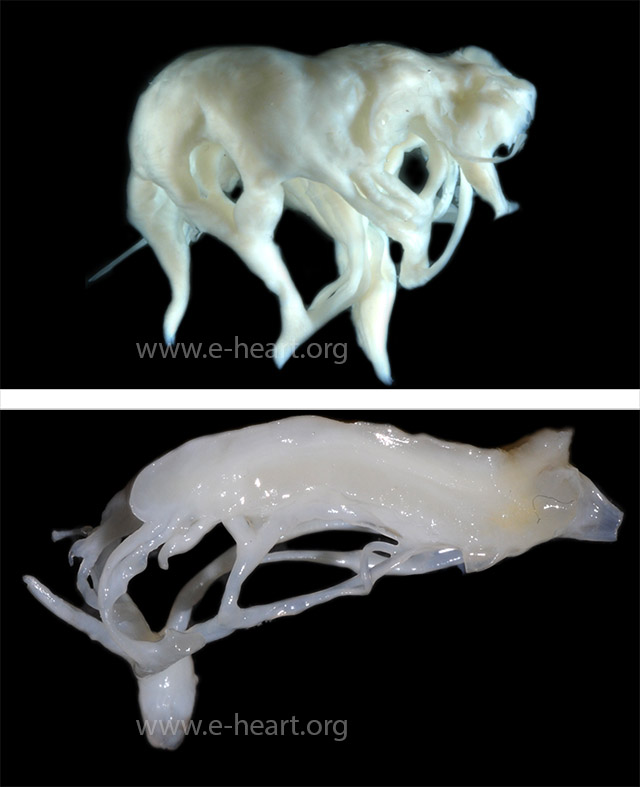 Myxomatous degeneration of the mitral valve produces distinct changes on the leaflet architecture. The atrial surface shows from slight bumps which protrude into the atrial cavity to frank billowing. These changes are secondary to the massive infiltration of the spongiosa layer by mucopolysaccharides. These infiltration also affects the chordae tendineae producing irregular thickening which shows a “beaded” appearance of the chordae. This should not be confused with chordal fusion. The lower image shows a section perpendicular to the atrialis layer. The infiltration of the spongiosa by mucopolysaccharides imparts a gelatinous appearance to the leaflet. The chordae show the distinct irregular thickening.
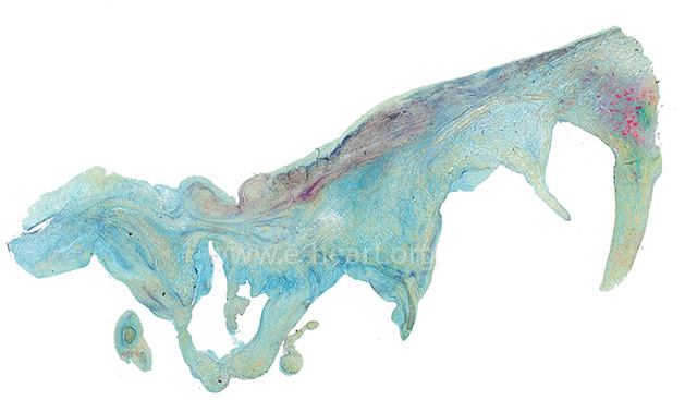 Microscopic examination of a Movat stain shows the massive infiltration of the spongiosa layer by mucopolysaccharides (green). The atrialis layer shows prominent elastosis (black elastic lamellae). The chordae tendineae show that the dense fibrous tissue (yellow) is infiltrated also by mucopolysaccharides.