

Myocardial interstitial fibrosis II
 Light microscopy of endomyocardial biopsy in restrictive cardiomyopathy.
A. Myocardium showing moderate hypertrophy with myocytes measuring between 30-40 micrometers in average diameter. There is no evidence of storage material in the myocytes sarcoplasm. The endocardium is thickened and the interstitial space is increase by eosinophilic connective tissue material. (Hematoxylin and eosin, X 100).
B. Connective tissue stain (trichrome) show dense blue material (fibrosis) which thickens the endocardium and expands the interstitial space surrounding practically every myocyte in the biopsy sample. This forms a restrictive girdle that restricts contractility (contraction and relaxation) of the myocytes. (Masson Trichrome stain, X100).
C. The fibrous tissue stains yellow in this stain with the same pattern as shown in B. However in this stain there is also accumulation of elastic lamellae in the endocardium consistent with endocardial fibroelastosis in addition to the interstitial fibrosis. (Movat pentachrome, X 100).
D. This illustration highlights the contours of the myocytes with interstitial fibrous tissue as well as the thickened endocardium in the upper and lower surfaces of the biopsy piece. (Masson trichrome, X100)
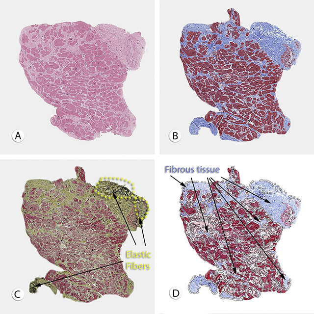
Light microscopy of endomyocardial biopsy in restrictive cardiomyopathy.
A. Myocardium showing moderate hypertrophy with myocytes measuring between 30-40 micrometers in average diameter. There is no evidence of storage material in the myocytes sarcoplasm. The endocardium is thickened and the interstitial space is increase by eosinophilic connective tissue material. (Hematoxylin and eosin, X 100).
B. Connective tissue stain (trichrome) show dense blue material (fibrosis) which thickens the endocardium and expands the interstitial space surrounding practically every myocyte in the biopsy sample. This forms a restrictive girdle that restricts contractility (contraction and relaxation) of the myocytes. (Masson Trichrome stain, X100).
C. The fibrous tissue stains yellow in this stain with the same pattern as shown in B. However in this stain there is also accumulation of elastic lamellae in the endocardium consistent with endocardial fibroelastosis in addition to the interstitial fibrosis. (Movat pentachrome, X 100).
D. This illustration highlights the contours of the myocytes with interstitial fibrous tissue as well as the thickened endocardium in the upper and lower surfaces of the biopsy piece. (Masson trichrome, X100)