

Cardiac Amyloidosis - XIV
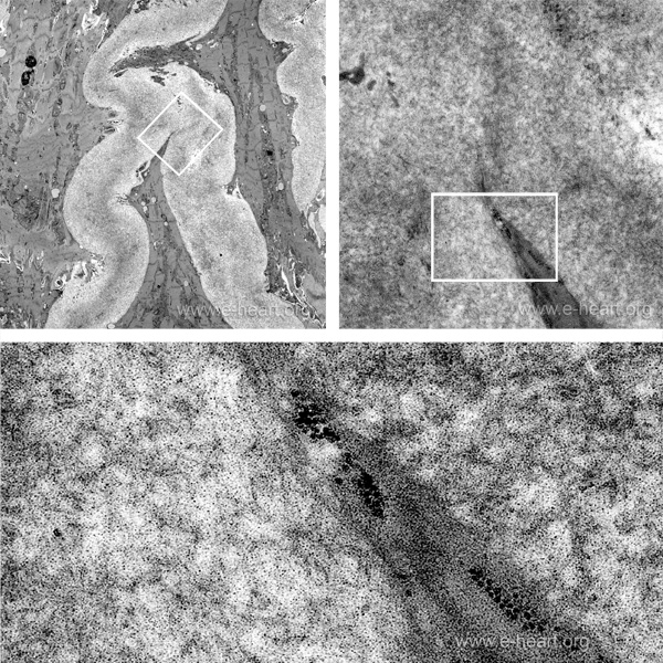
This is a transmission electron micrograph of and endomyocardial biopsy showing perimyocytic deposition of amyloid. The amyloid fibrils, measure and average of 10 nanometers in diameter and form an electron dense deposit. The upper right image shows several cardiac myocytes surrounded by amyloid. The white rectangles show areas of close up which is in turn shown at higher magnification. The 10 nm amyloid fibrils are disctinc thread-like structures.