

Atrioventricular Valves
 Atrioventricular valves.
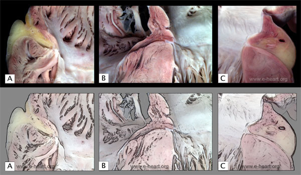
A. Close-up view of a coronal section through the right lateral border of the right atrium and ventricle. The pectinate muscles of the atrium are prominent. The atrioventricular sulcus is deep and contains adipose tissue. A cross section of the right coronary artery cruising through is also shown in the sulcus. At the junction of the right atrium with the right ventricle there is a prominent fibrous annulus to which the tricuspid valve is anchored. B. Close up view of the atrial septum as it merges with the interventricular septum. The anchoring sites for the tricuspid and mitral valves are evident. C. Close up view of the lateral border of the left atrium as it joins the left ventricle shows the atrioventricular sulcus. The adipose tissue in the sulcus surrounds the left circumflex coronary artery and the great cardiac vein. The mitral fibrous annulus is a discrete fibrous structure joining the atrial and the ventricular muscle.
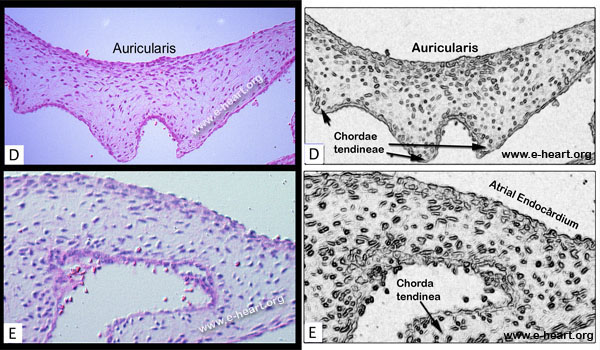 D. Light micrograph of the mitral valve of a fetus, showing a somewhat myxoid stroma without differentiation into the layered structure of the adult valve. The atrial side of the valve is indicated (auricularis) ( H&E , 50 X). E. Histologic section of the valve shows plump cells embedded in the myxoid matrix. Note the single layer of endothelial cells lining the surface of the valve. F. Scanning electron micrograph showing a mosaic-like pattern of arrangement of the endothelial cells. A prominent bulge corresponds to the nucleus in the central portion of the cell. Small, scant microvilli are present. ( 2,500 X)
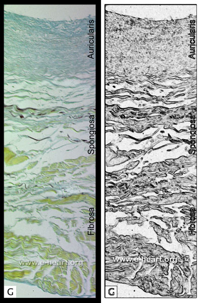 G. Low magnification micrograph of a section of the mitral valve. The layers of the atrioventricular valves vary slightly according to the region (whether basal or distal) of the leaflet. The fibrosa is the most consistent layer. It is formed by collagenous (yellow) bundles. The spongiosa contains loosely arranged connective tissue, proteoglycans and small amounts of elastic fibers. It is most prominent in the basal portion of the valve. The auricularis is also rich in proteoglycans and is located on the atrial side of the proximal portions of the valve (not shown) (Movat pentachrome, 25X).
Back to Cardiac Structure