Cardiac Allograft Vasculopathy (V)
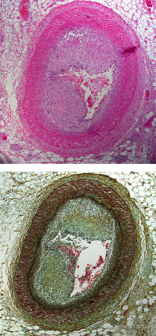
 Microscopic examination of allograft vasculopathy lesions shows concentric intimal proliferation composed of smooth muscle cells and less-differentiated spindled cells (myofibroblasts or ''myointimal'' cells) There is accompanying abundant deposition of proteoglycans which stain blue in the Movat stain (lower images). These areas are the equivalent of the white neointima seen on gross examination. There is a distinctly different staining pattern and distribution, compared with conventional atherosclerosis, and more similar to angioplasty-related restenotic lesions. The panel below shows the cicumflex coronary artery of a patient with minimal vasculopathy, but, the artery supplying the atrial myocardium shows sever vasculopathy (black rectangle)
At higher magnification this example shows an eccentric plaque showing the proliferative intima in the right half of an epicardial coronary artery. (H&E, X10). B. The proliferative intima in this case is formed of mature extracellular matrix rich in glycosaminoglycans (green). The internal elastic lamina (black) is intact, and the media of the vessel is also intact (dark red). (Movat pentachrome, X10).