

Antibody Mediated Rejection (AMR) - I
Antibody-mediated rejection (AMR) is an immunopathologic process in which the humoral arm of the immune response produces antibodies against epitopes in the graft. These antibodies in turn, activate the complement system and the complement activation results in injury to the graft. This type of rejection is poorly responsive to conventional immunosuppression, which targets the cellular arm of the immune response. Old terminology such as vascular rejection, microvascular rejection, and humoral rejection should be avoided as it has only led to confusion in the literature. The preferred terminology in the ISHLT-WF2004 is AMR.
The histologic features that allow for the identification of this type of rejection on endomyocardial biopsies as defined in the ISHLT-WF2004 and its companion article on AMR include: ‘‘capillary endothelial changes (swelling or denudation with congestion), macrophages in capillaries, neutrophils in capillaries, interstitial edema and/or hemorrhage and fibrin in vessels.’’ If these features are observed in the biopsy and there is unexplained cardiac dysfunction, the revised working formulation proposed that immunofluorescence or immunohistochemistry, in the absence of frozen tissue, be performed. Immunopathologic evidence of AMR include ‘‘—Immunoglobulin (IgG, IgM and/or IgA) plus complement deposition (C3d, C4d and/or C1q) in capillaries by immunofluorescence on frozen sections; and/or —CD68 staining of macrophages within capillaries (CD31- or CD34-positive) by immunohistochemistry; and —C4d staining of capillaries by paraffin immunohistochemistry.’’
Examples of the capillary pattern of complement deposition
are shown in in the images below. The image on the left is a stain for C4d deposits, which shows strong flurescent signal in capillary patters. The image on the right is stained for C3d. It is also recommended that these patients undergo assessment
for circulating antibodies to HLA class I or II as
well as non-HLA donor antigens. An EMB with no histologic
or immunopathologic evidence of AMR is graded
0 (AMR 0). If the immunofluorescence or immunohistochemical
staining supports the histologic features of AMR,
the biopsy is considered positive (AMR 1). 
This micrograph shows the comparison of immunofluorescence stainig for C4d (top) and immunoperoxidase staining for C4d (below).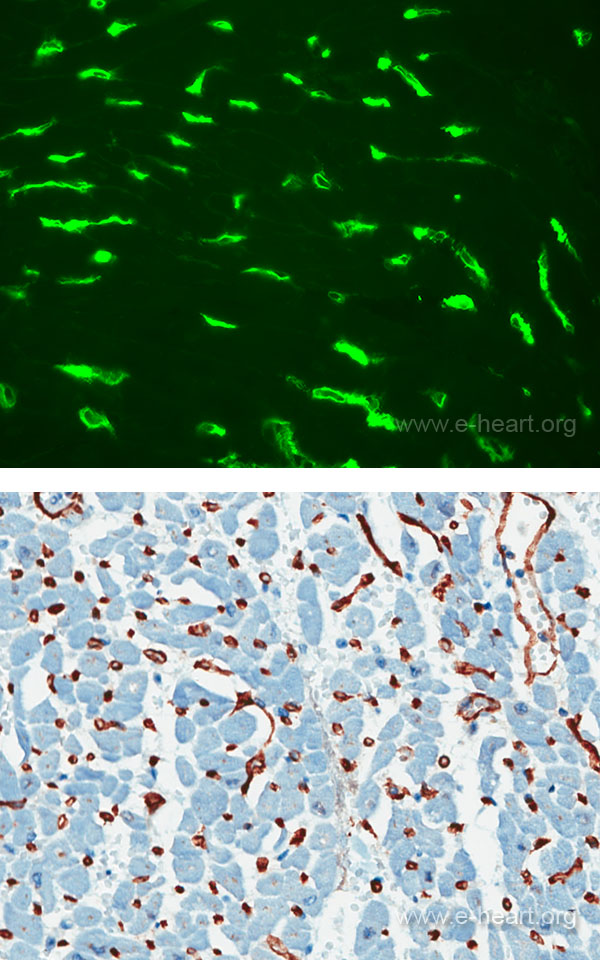
In this image the antibodies used are anti-C3d using immunofluorescence (top) and immunoperoxidase staining methods (bottom)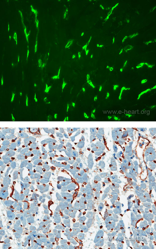
Regulators of complement activation are also an important part in the controls of the humoral response to the allograft.