

Cardiac Structure - Right Atrium
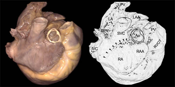 This is a right-lateral view of the right atrium (RA) the right atrial appendge (RAA) the superior vena cava (SVC) and the inferior vena cava (SVC). There is a groove present between the superior vena cava and the right atrium which is called the "sulcus terminalis" and is demaractated by the arrowheads in the illustraion. The outflow tract of the right ventricle (RVOT) is anterior to the tip of the right atrial appendage (RAA). The root of the aorta (Ao) is posterior to the root of the pulmonary artery (PA). A cephalad view of the fetal left and right atrial appendages shows diferences in the contours of the atrial appendage lumina.
 Cephalad view of the right atrial appendage. The pectinate muscles (PM) are seen forming parallel bundles that run in a cephalad direction to merge with a broad band of muscle which is the crista terminalis (dashed area). The sinus venarum (SVe) is the portion of the atrial wall located between the openings of the superior vena cava and the inferior vena cava.
In addition, a medial view of the right side of the atrial septum reveals other anatomic landmarks.
Cephalad view of the right atrial appendage. The pectinate muscles (PM) are seen forming parallel bundles that run in a cephalad direction to merge with a broad band of muscle which is the crista terminalis (dashed area). The sinus venarum (SVe) is the portion of the atrial wall located between the openings of the superior vena cava and the inferior vena cava.
In addition, a medial view of the right side of the atrial septum reveals other anatomic landmarks.